


| 1. | |
Liver-Fas antibody-induced acute liver injury | ● Fas antigen is important in programmed cell death in the liver [Ogasawara J et al. Nature 364:806 (1993).] ● Loss of Caspase-8 Protects Mice Against Inflammation-Related Hepatocarcinogenesis but Induces Non-Apoptotic Liver Injury [Liedtke C et al. Gastroenterology 141:2176 (2011)]. |  |
|
Liver-acetaminophen-induced acute liver injury | Acetaminophen (APAP) overdose leads to drug-induced liver damage. ● C-Jun N-terminal kinase (JNK) plays a major role to promote APAP-induced hepatotoxicity [Gunawan BK et al. Gastroenterology 131:165 (2006)]. ● Deletion of apoptosis signal-regulating kinase 1 (ASK1) attenuates APAP-induced liver injury by inhibiting JNK. ASK1 likely is involved in the liver damage by prolongation of JNK activation [Nakagawa H et al. Gastroenterology 135:1311 (2008)]. | |
|
| 2. | |
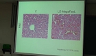
Liver-steatohepatitis | ● Loss of B-catenin in the liver leads to defective cholesterol and bile acid metabolism in the liver and increased susceptibility to developing steatohepatitis in the face of metabolic stress [Behari J et al. Am J Pathol 176:744 (2010)]. | |
|
Liver-fibrosis-angiopoietin 1 | ● Hepatic stellate cells secrete angiopoietin 1 that induces angiogenesis in liver fibrosis [Taura K et al. Gastroenterology 135:1729 (2008)]. | |
|
| 3. | |
Liver-fibrosis-angiotensin II | ● Angiotensin II activates IkB Kinase Phosphorylation of RelA at Ser536 to promote myofibroblast survival and liver fibrosis [Oakley F et al. Gastroenterology 136:2334 (2009)] | |
|
Pancreas-Shh-induced metaplasia in chronic pancretitis | ● Pancreatic duct glands are distinct ductal compartments that react to chronic injury and mediate Shh-induced metaplasia [Strobel O et al. Gastroenterology 138:1166 (2010)] ● This study characterizes ths novel ductral compartment that is gathered in outpouches, pancreatic duct glands (PDG). ● PDG retain expression of gastric mucins and Shh. ● In response to chronic injury, PDG up-regulate developmental genes and proliferative rates and undergo an Shh-mediated mucinous metaplasia. | |
|
| 4. | |
Pancreas-Hedgehog signaling in acinar cell regeneration | ● Hedgehog Signaling Is Required for Effective Regeneration of Exocrine Pancreas [Fendrich V et al. Gastroenterology 135:621 (2008)] ● Using a model of cerulein-mediated injury and repair, this study demonstrates that mature exocrine cells actively contribute to regenerating pancreatic epithelium through formation of metaplastic ductal intermediates. ● Acinar cell regeneration is associated with activation of Hedgehog (Hh) signaling. | |
|
Pancreas-Hedgehog signaling in β-cell dedifferentiation | ● Elevated Hedgehog/Gli signaling causes β-cell dedifferentiation in mice [Landsman L et al. PNAS 108:17010 (2011)] ● Increased Hh signaling leads to impaired β-cell function and insulin secretion, resulting in glucose intolerance in transgenic mice. ● Sustained high Hh levels in some insulin-producing cells further eroded the β-cell identity and eventually led to the development of undifferentiated pancreatic tumors. | |
|
| 5. | |
Pancreas-Notch signaling in a mouse model of pancreatic ductal adenocarcinoma | ● Inhibition of r-secretase activity inhibits tumor progression in a mouse model of pancreatic ductal adenocarcinoma [Plentz R et al. Gastroenterology 136:1741 (2009)] ● Expression of activated Kras (KrasGOF) in acinar cells leads to PanIN formation, which is accelerated in the presence of activated Notch. ● Loss of the tumor suppressor p53 (p53LOF) allows progression to invasive tumors, which can be repressed by blocking Notch signaling with a GSI. | |
|
Pancreas-Notch signaling in acinar-to-B-cell conversion | ● Notch signaling as gatekeeper of rat acinar-to-B-cell conversion in vitro [Baeyens L et al. Gastroenterology 136:1750 (2009)] ● Ductal metaplasia of cultured acinar cells can be directly induced by Notch, or indirectly (still requiring Notch function) by epidermal growth factor (EGF) receptor agonists. ● Cultured acinar cells can be efficiently forced to become functional B-cells, in the presence of EGF and leukemia inhibitory factor (LIF), by an initial activation of Notch (acinar-to-duct) followed by its inhibition (duct-to-B-cell). | |
|
| 6. | |
Intestine-NOD2 null mice | ● Nod2-dependent regulation of innate and adaptive immunity in the intestinal tract [Kobayashi KS et al. Science 307:731 (2005) ] ● Protective immunity mediated by Nod2 recognition of bacterial muramyl dipeptide is abolished in Nod2-deficient mice. ● These animals are susceptible to bacterial infection via the oral route but not through intravenous or peritoneal delivery. ● Nod2 is required for the expression of a subgroup of intestinal anti-microbial peptides, known as cryptdins. | |
|
Intestine-NOD2 2939iC mice | ● Nod2 mutation in Crohn’s disease potentiates NF-kB activity and IL-1b processing [Maeda S et al. Science 307:734 (2005)] ● NOD2 2939iC mice exhibited elevated NF-kB activation in response to MDP and more efficient processing and secretion of the cytokine interleukin-1b (IL-1b). ● These effects are linked to increased susceptibility to bacterial-induced intestinal inflammation and identify NOD2 as a positive regulator of NF-kB activation and IL-1b secretion. | |
|
| 7. | |
Intestine-MyD88 null mice | ● Recognition of commensal microflora by Toll-Like Receptors is required for intestinal homeostasis [Rakoff-Nahoum et al. Cell 118:229 (2004)] ● Severe susceptibility to colonic injury in MyD88-deficient mice. ● TLR-mediated recognition of commensals in the colon regulates production of tissue protective factors. ● Commensal bacteria are recognized by TLRs under normal steady-state conditions, and this interaction plays a crucial role in the maintenance of intestinal epithelial homeostasis. | |
|
Intestine-TLR5 | ● Pathophysiological role of Toll-like receptor 5 engagement by bacterial flagellin in colonic inflammation [Rhee SH et al. PNAS 102:13610 (2005)] ● Flagellin TLR5 response is restricted to the basolateral apect of colonic mucosa. ● Flagellin exposure to the injured colonic mucosa induces in vivo activation of MEK1/2 in colonic mucosa. ● Flagellin exposed to injured colonic mucosa significantly aggravates the clinical symptoms of colitis. ● Flagellin exposure to injured colon causes apoptosis in colonic epithelium. | |
|
| 8. | |
Intestine-EP2-PGE2 | ● Acceleration of intestinal polyposis through prostaglandin receptor EP2 in ApcΔ716 knockout mice [Sonoshita M et al. Nat Med 7:1048 (2001)] ● Homozygous deletion of the gene encoding a cell-surface receptor of PGE2, EP2, causes decreases in number and size of intestinal polyps in ApcΔ716 mice (a mouse model for human familial adenomatous polyposis). This effect is similar to that of COX-2 gene disruption. | |
|
Intestine-EP2-PGE2 | ● Catellone MD et al. Science 310:1504 (2005) | |
|
| 9. | |
Intestine-MUC2 | ● Muc2-Deficient Mice Spontaneously Develop Colitis, Indicating That Muc2 Is Critical for Colonic Protection [Van der Sluis M et al Gastroenterology 131:117 (2006)] ● Muc2 -/- mice showed clinical signs of colitis (as of 5 weeks), aggravating as the mice aged. Microscopic analysis of the colon of Muc2 / mice showed mucosal thickening, increased proliferation, and superficial erosions. | |
|
Lung-KRAS | ● Wildtype Kras2 can inhibit lung carcinogenesis in mice [Zhang Z et al. Nat Genet 29:25 (2001)] ● Mice with a heterozygous Kras2 deficiency were highly susceptible to the chemical induction of lung tumors when compared to wildtype mice. ● Wildtype Kras2 has tumor suppressor activity and is frequently lost during lung tumor progression. | |
|
| 10. | |
Lung-PTEN | ● Pten controls lung morphogenesis, bronchioalveolar stem cells, and onset of lung adenocarcinomas in mice [Yanagi S et al. J Clin Invest. 117:2929 (2007)] ● Generated mice deficient for Pten specifically in bronchioalveolar epithelial cells (a) 90% died within 2 hours of birth (b) Surviving mice developed spontaneous lung adenocarcinomas (c) Hyperplasia of bronchioalveolar epithelial cells and myofibroblast precursors, enlarged alveolar epithelial cells, and impaired production of surfactant proteins (d) Increased numbers of bronchioalveolar stem cells (BASC) ● Pten is essential for both normal lung morphogenesis and the prevention of lung carcinogenesis, possibly because this tumor suppressor is required for BASC homeostasis | |
| 10. | |
Lung-NFkB | ● Requirement for NF-kB signalling in a mouse model of lung adenocarcinoma [Meylan E et al. Nature, 462:104 (2009)] ● Concomitant loss of p53 (also known as Trp53) and expression of oncogenic Kras(G12D) resulted in NF-kB activation in primary mouse embryonic fibroblasts ● Inhibition of NF-kB signalling induced apoptosis in p53-null lung cancer cell lines ● Inhibition of NF-kB pathway in lung tumours in vivo resulted in significantly reduced tumour development ● Together, these results indicate a critical function for NF-kB signalling in lung tumour development and, further, that this requirement depends on p53 status | |
| 11. | |
Lung-NKX2-1 | ● Suppression of lung adenocarcinoma progression by Nkx2-1 [Winslow MM et al. Nature, 473:101 (2011)] ● Nkx2-1 negativity is pathognomonic of high-grade poorly differentiated tumours. ● Nkx2-1 controls tumour differentiation and limitsmetastatic potential in vivo. ● Nkx2-1 constrains tumours in part by repressing the embryonically restricted chromatin regulator Hmga2. ● Data specifically link Nkx2-1 downregulation to loss of differentiation, enhanced tumour seeding ability and increased metastatic proclivity. | |

 대구광역시 동구 동내로 64(동내동 1119) 우)41061
COPYRIGHT keris. all rights reserved